KOKorean
 ENEnglish
 JP日本語
JP日本語 CN中文(简体)
 TW中文(繁體)

Breast Filler and Injected Material Removal in Seoul: When to Remove It, Breast Screening and How the Space Is Restored
If lumps, pain, a change in shape or inflammation appear, removal is usually considered. Even without symptoms, it is worth agreeing in advance how your breasts will be screened. Material injected into the breast often scatters across several layers, from under the skin to beneath the muscle, so some may remain even after surgery. The breast becomes smaller in proportion to what is removed and may look loose. So when to restore it with your own fat is decided once inflammation has settled and the tissue is stable. If you are considering treatment in Seoul, you can send photos, your injection history and any breast imaging reports first.
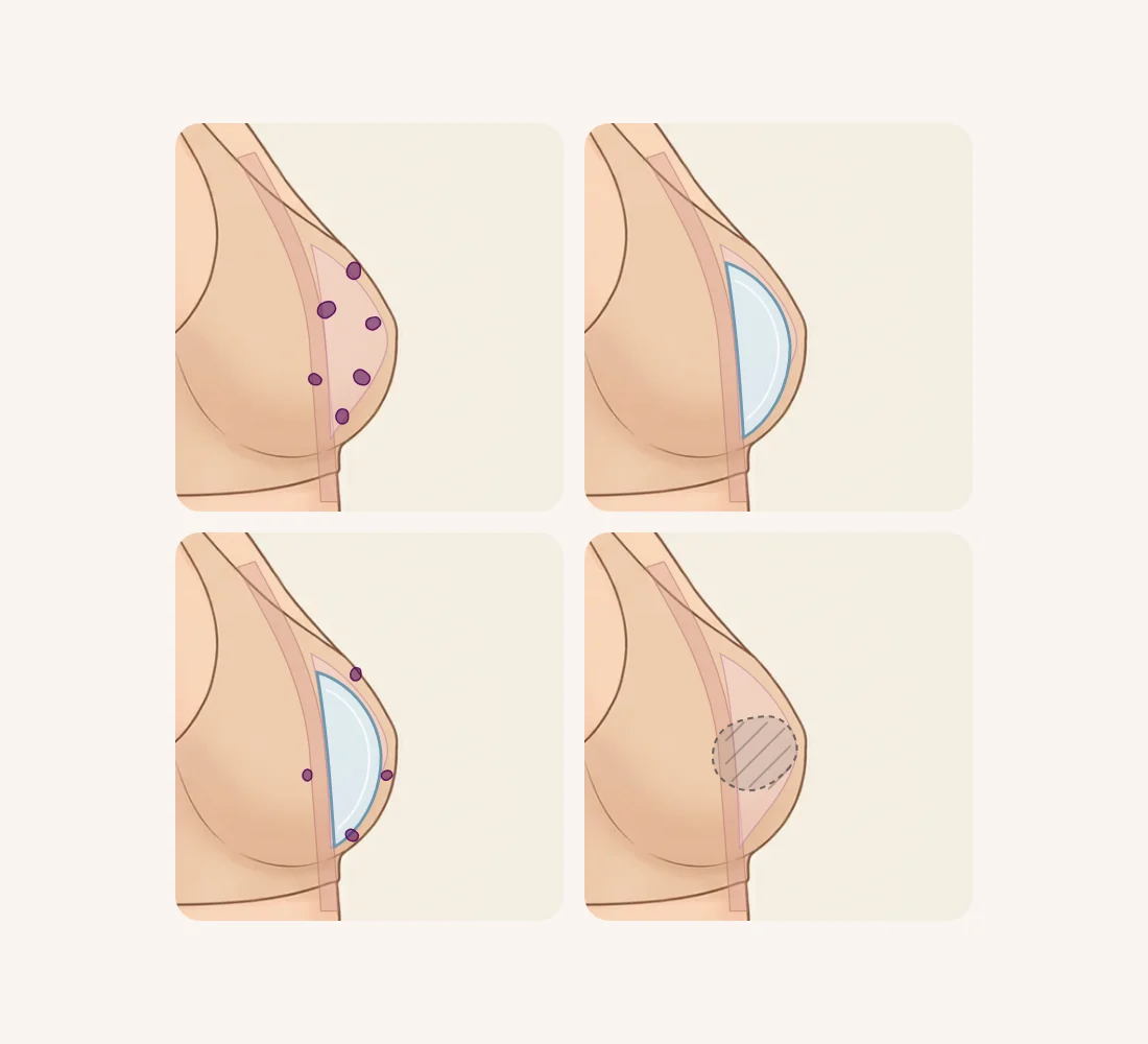
What is the first thing to work out: an implant, or injected material?
The first thing to establish is what is actually in the breast. A silicone implant is a pouch with a shell around its contents, so it can be removed or exchanged as one piece. Injected material has no shell. It spreads directly between fat, the glandular tissue (the tissue that makes milk) and muscle. Over time the body often wraps it in a hardened layer of scar-like tissue, called a capsule. Both may be called "breast foreign material", but they are removed differently and leave different results.
What has been injected into breasts has changed over the decades. Paraffin was used in the early twentieth century and liquid silicone later. From the late 1980s, polyacrylamide hydrogel (a gel that holds a lot of water) was used widely in Ukraine, Russia and China.1 Today people also come with problems after hydrogel fillers such as Aquafilling or hyaluronic acid injected into the breasts, and many were injected abroad and do not know the product. A Japanese study, from a country where silicone and paraffin injections are banned, noted that some patients injected overseas or injected several times did not know what they had received.5
What is in the breast?
1Injected material only
Removal and filling of the space are planned together
The case covered in this guide
2A silicone implant only
Implant exchange or removal is considered
See revision breast surgery
3Injected material plus an implant
Each is mapped on imaging, then the order of treatment is set
Plan the order first
4Not sure what it is
Ultrasound or MRI comes first, along with old records and what you were told
Imaging first
Implant exchange or removal, and filling with your own tissue after implant removal, are covered on our Revision Breast Surgery page. This guide focuses on injected fillers and foreign material.
What problems can injected material cause?
Each material shows itself at a different time and in a different way.
- Paraffin: it often looked acceptable at first, but records describe hard knots and draining tracts (fistulae, small channels that leak pus) appearing five or ten years later.1
- Liquid silicone: the average time from injection to complications is about nine years. Painful lumps appear within two or three years in some women, and only after ten or fifteen years in others.1
When silicone works its way into the skin, circulation in the skin can get worse in stages. It starts with fine visible capillaries and, at worst, ends in skin necrosis (skin tissue dying). In one reported case, much of the silicone injected into the breasts had moved down to the abdomen over thirty years.1
With polyacrylamide hydrogel, migration, lumps, pain, infection, firmness and disfigurement have appeared from a few months to three years after injection. Leakage of the gel back along the needle track is thought to be one cause of migration.1 In China, 161 of 183 adverse event reports received between 2002 and November 2005 involved breast injections, and the production, sale and use of the material were then banned.1 A study of 135 women who had this gel removed reported these problems: hard lumps and nodules (60.0%), pain (27.4%), anxiety (22.2%) and migration (13.3%). Others were asymmetry (8.1%), swelling (5.9%), infection (5.2%) and calcification (3.7%).2 Put simply, about six in ten of the women who had it removed had firm lumps.
Even hyaluronic acid, which the body can break down, frequently forms multiple encapsulated lumps when injected into the breasts. Once encapsulated, these lumps often do not dissolve with enzyme injections and have had to be removed surgically.5
How does injected material affect breast screening?
Screening matters as much as shape. In the 135-patient hydrogel study, breast cancer was found in 4 women (3.0%). The authors stressed that, although rare, it can be hidden by the gel and the tissue around it.2 In another series, two candidates whose pre-operative tests strongly suggested cancer were referred for diagnosis and excluded from fat grafting.4
On mammography (a breast X-ray), a silicone-injected breast may show many round masses of 0.2 to 2.0 cm, often calcified, or broad white areas when large volumes were used.1 In other words, the material can block the view of what lies behind it. A radiology review therefore notes that mammography remains the screening test, but that breast MRI without contrast dye is a useful extra test for women with injected material. MRI with contrast dye (a medicine that makes images clearer) is needed when infection or cancer is suspected.6 That is why you should always tell the screening center what was injected and when.
Which layers of the breast does the material occupy?
Beneath the skin of the breast lie the fat layer, the glandular tissue, a thin space behind the gland, and the large chest muscle (pectoralis major). Injected material rarely stays in one layer. In one reported case, MRI showed it both behind the gland and beneath the muscle.1 When the area is refilled after removal, fat has been placed in several layers around the cavity rather than into the empty space itself.4
Which layer the material occupies changes both the method and the chance that some is left. A study of 436 breasts that had hydrogel removed linked four things to more complications after surgery:
- Gel that had soaked into the glandular tissue on MRI
- A longer time since injection
- Previous needle aspiration (drawing material out with a syringe)
- A hardened capsule3
How is it removed, and how far can removal go?
The usual method depends on the state of the material.
| Situation | Common approach | Good to know |
|---|---|---|
| Hydrogel pooled in a large pocket | Expressing and rinsing it out through an incision, cannula suction | Harder to clear when injected in many small sites1 |
| A hardened capsule around the material | Removing the capsule with the material | A capsule left behind keeps the cavity from closing4 |
| Encapsulated hyaluronic acid lumps | Excision, for example from the edge of the areola | May not dissolve with enzyme injections5 |
| Silicone or paraffin tangled in the gland | Gland resection may be needed | Small incisions are often not enough5 |
| Silicone with skin breakdown and fistulae | Wide excision, up to mastectomy | Usually chosen in advanced cases1 |
A cannula is a thin tube with a blunt tip, and the areola is the darker skin around the nipple. Hydrogel comes out much more completely from a large pocket than from many small scattered sites.1 Even so, in the 135-patient study, gel was left behind after removal in 17.0% and loose skin was seen in 7.4%.2 The breast becomes smaller in proportion to what is removed, and skin that has been stretched for years may look loose.
When, and with what, is the space restored?
Leaving the breast unfilled lowers the chance of further surgery, but can lower satisfaction. A 20-year comparison of 436 breasts found:
- Complications after surgery: 50.0% with same-day reconstruction, 31.3% with delayed reconstruction and 26.7% with no reconstruction
- Reoperation: 26.2%, 18.8% and 13.0% in the same order
- Satisfaction: the no-reconstruction group scored lower for satisfaction with the breasts and for psychosocial and sexual well-being than either reconstruction group.3
The authors concluded that delayed reconstruction is a safe and effective option.3 Delayed filling had fewer complications than same-day filling, and higher satisfaction than no filling.
A series of 162 women who had fat grafted on the same day as removal recovered without severe complications. But it excluded anyone with infection or skin ulceration (an open sore) from the start, and stated that fat grafting should wait until any infection is fully controlled. Fat was placed over an average of 1.6 sessions (range 1 to 3).4 So whether same-day filling is possible depends on the absence of inflammation and on the condition of the tissue.
How we approach it at Bonita Clinic
At Bonita Clinic, we perform breast foreign material removal as surgery under general anesthesia or sedation, with same-day discharge or a one-night stay, and stitches.
Treatment options you can discuss in Seoul
Bonita Clinic has worked from the same place since 1995, and Dr. Kwang-Seop Chung, a board-certified plastic surgeon (Brazil), sees each patient in person. For injected breast material, consultation covers:
- Ultrasound to locate the remaining material, its size and the state of the tissue
- Surgical removal by hand or fine cannula, freeing adhesions while limiting damage
- Fat grafting with your own fat to fill the space, timed to the tissue's condition
- Combined laser treatment for marks and skin texture, and follow-up of scars at our Scar Clinic
- Separate planning through our revision breast surgery service if an implant is also present
Breast MRI, mammography and other screening tests mentioned here are general medical information. Bring any reports you have; whether further imaging is needed is discussed after the examination.
Why do the plan and cost differ from person to person?
The name of the problem says little about the operation. The amount of work does.
- How much was injected, and into which layers
- Whether a hardened capsule must come out too
- Fat grafting the same day, later, or not at all
- General anesthesia or sedation, overnight stay
- An implant that also needs attention
- Infection or skin damage to settle first
Final plan and quote after an in-person exam and ultrasound
The final estimate is given only after the in-person examination, once the layers involved, the need for capsule removal and the restoration plan are clear.
What can photos show, and what needs an in-person exam?
Photos show the outside of the problem. The decisions depend on what lies beneath.
Photos can show
- Size difference and shape changes
- Visible lumps or bulges
- Skin color, thinning or capillaries
- How the shape changed (older photos)
Needs an in-person exam
- Which layers hold the material
- Whether gel has entered the gland
- Capsule hardness and adhesion
- Active infection
- How much fat is available to graft
Helpful photos: front, both sides at 45 degrees and side views, arms relaxed and then raised, in good light, without your face. Photos from before the first injection help us understand your original shape. Please also keep any mammography, ultrasound or MRI reports ready. You can message us the photos together with whatever you recall about the injections and any screening results.
Planning your stay and recovery
The confirmed details are those above: surgery under general anesthesia or sedation, same-day discharge or one night in the clinic, and stitches. How long to stay in Seoul, how many follow-up visits you need and when stitches come out all vary. They depend on how much is removed and whether fat is grafted at the same time. These are planned with you at consultation.
Recovery after breast material removal usually goes like this:
- After surgery: swelling and bruising are managed with compression and cold packs.
- As the swelling settles: the contour of the breast becomes clearer.
- Settling: touch-ups are judged only after the removal and grafted areas have stabilized.
After you return home, seek care promptly if redness, heat, pain, fever or discharge increases. For future screening, tell the radiologist that material was injected and later removed, and whether any may remain; bringing the operation summary helps them read your images.
Real treatment cases
These cases appear on Bonita Clinic's breast revision page. Results vary from person to person, and both show how volume decreases in proportion to what is removed.
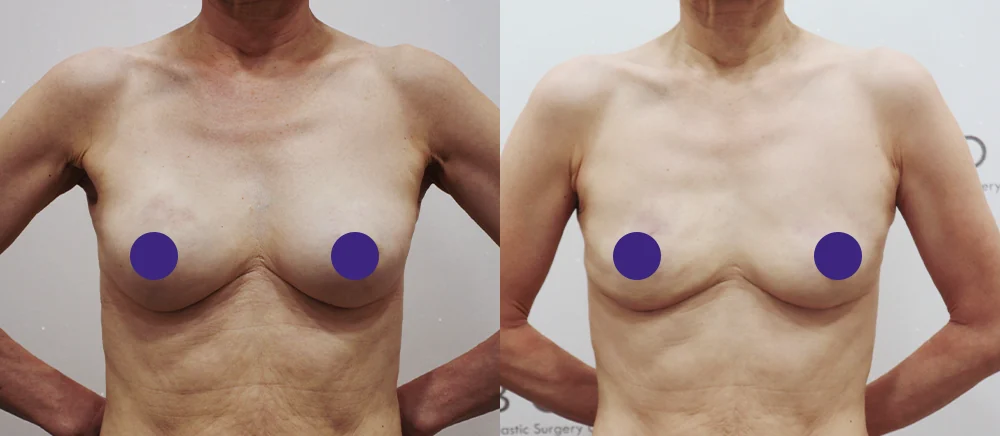
Both breasts rounded and full up to the upper pole
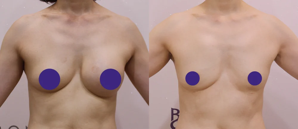
Both breasts enlarged, with a difference in shape between sides
* Results vary from person to person. Side effects are possible, so please decide after a thorough consultation with a doctor.
Frequently asked questions
I have no symptoms right now. Should it still come out?
Whether surgery is needed now varies from person to person. Injected material can make breast screening harder, so a screening plan should be agreed in advance.6 If lumps, pain or a change in shape appear, it is better not to wait. We look at the current state with ultrasound and decide together.
How much smaller will my breasts be after removal?
It depends on how much was injected and how much glandular tissue you have. Removing the material together with the hardened capsule reduces volume accordingly, and skin that was stretched may look loose. That is why removal and restoration are planned together.
Can fat be grafted on the same day as removal?
Sometimes, but conditions apply. The same-day series excluded patients with infection or skin ulceration,4 and another study found more complications with same-day reconstruction than with delayed reconstruction.3 Timing depends on whether inflammation has settled and whether gel has entered the glandular tissue.
Will it affect breastfeeding or future screening?
Injected hydrogel itself has been reported to cause breastfeeding difficulties.4 The effect of removal also depends on how much the surgery involves the gland, so tell us if you plan a pregnancy. For screening, sharing both your injection history and your removal history helps the radiologist.6
Where will the scars be?
Along the incisions, for example at the edge of the areola or in the fold under the breast.5 Silicone or paraffin tangled in the gland may need larger incisions.5 We keep incisions as small as possible and place them where they show least, and remaining scars are followed up at our Scar Clinic.
Before your consultation
- When, where (including which country), how many times and roughly how much was injected on each side, and any product name you were told
- Whether material or pus was ever drawn out with a needle; previous aspiration has been linked to later complications3
- Dates and reports of mammography, ultrasound or MRI
- Any breast implant surgery or fat grafting in the past
- Pregnancy or breastfeeding plans, and any family history of breast cancer
Bonita Clinic is on the 3rd floor of 565 Gangnam-daero, Seocho-gu, Seoul, about 100 m on foot from Exit 8 of Nonhyeon Station (Line 7). Read more on our Breast Surgery Revision page. For implant exchange or removal see Revision Breast Surgery. For material injected into the buttocks, hips or legs see Buttock & Leg Surgery Revision and the guide to buttock and leg filler removal.
References (6)
Sources that support the explanations in this article. Study findings describe general trends and do not guarantee individual results.
- Peters W, Fornasier V. Complications from injectable materials used for breast augmentation. Can J Plast Surg. 2009;17(3):89-96.doi:10.1177/229255030901700305Historical review of breast injectables (paraffin, silicone, polyacrylamide hydrogel): time to complications, skin involvement and migration, mammographic patterns, easier removal from large pockets, mastectomy for advanced silicone granulomatosis, the ban in China
- Guan D, Yuan H, Sun K, et al. Clinical Experience in the Management of the Polyacrylamide Hydrogel (PAAG) Associated Complications Including Four Breast Cancer Cases: A Retrospective Study of 135 Cases. Aesthetic Plast Surg. 2025;49(3):779-787.doi:10.1007/s00266-024-04259-9Retrospective study (abstract) of 135 hydrogel removals: symptom frequencies, 4 breast cancers (3.0%) and the risk of the gel concealing cancer, 17.0% residual gel and 7.4% skin laxity after removal, better satisfaction with reconstruction
- Liang Y, Sun Q, Chu X, et al. Evaluating the safety, efficacy, and risk factors of different strategies for breast reconstruction following polyacrylamide hydrogel removal: A 20-year retrospective cohort study of 436 cases. Asian J Surg. 2025;48(1):311-322.doi:10.1016/j.asjsur.2024.09.13320-year cohort (abstract) of 436 breasts: complications and reoperation after same-day, delayed or no reconstruction, lower satisfaction without reconstruction, risk factors including gland infiltration and previous aspiration, delayed reconstruction judged safe
- Gao Q, Zhai P, Qi J, et al. Breast Augmentation with Autologous Fat Grafting Immediately after Removal of Polyacrylamide Hydrogel and Fibrotic Capsule in 162 Patients. Breast Care (Basel). 2022;17(4):377-384.doi:10.1159/000522616Series of 162 women with fat grafting immediately after hydrogel and capsule removal: infected or ulcerated cases excluded, fat grafting delayed when infection is present, capsule removal to close the cavity, fat in surrounding layers, 1.6 sessions on average
- Sakai S, Ishii N, Nakamura Y, et al. Complications and Surgical Treatment of Breast Augmentation Using Autologous Fat Transfer and Fillers. Plast Reconstr Surg Glob Open. 2021;9(8):e3734.doi:10.1097/GOX.0000000000003734Japanese series of 20 operations: encapsulated hyaluronic acid lumps resistant to enzyme injection, gland resection needed for silicone and paraffin, unknown products after injections abroad, areolar and inframammary incisions
- Noreña-Rengifo BD, Sanín-Ramírez MP, Adrada BE, et al. MRI for Evaluation of Complications of Breast Augmentation. Radiographics. 2022;42(4):929-946.doi:10.1148/rg.210096Radiology review (abstract): mammography remains the screening test, non-contrast breast MRI as a supplement with free injected material, contrast MRI when infection or cancer is suspected
About this article
This article separates general medical information from Bonita Clinic's own approach. Because the right decision depends on each person's condition, it is not a substitute for a consultation.