KOKorean
 ENEnglish
 JP日本語
JP日本語 CN中文(简体)
 TW中文(繁體)

Sebaceous Cyst and Lipoma Removal in Seoul: Why the Sac Matters and When to Remove a Lump
Not every lump has to be removed right away. But a cyst that keeps refilling, flares up again and again, or is growing in a visible place is worth removing. A sebaceous cyst tends to come back if only its contents are squeezed out, so the sac wall is removed with it. A lipoma is a lump of fat in a thin capsule and is taken out whole. A red, painful cyst is often left to settle before removal. A lump that is growing fast, is large or lies deep is checked with imaging first. If you are considering removal in Seoul, you can send photos and a short history of the lump first to see what may fit your case.
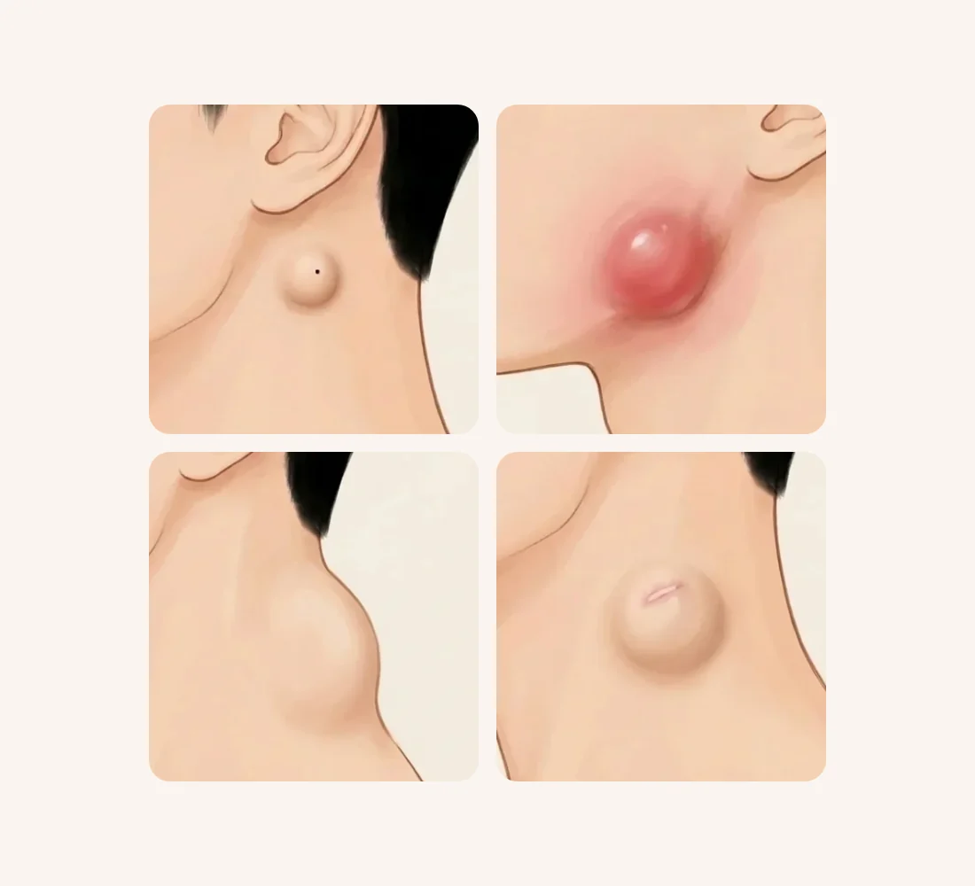
Where do sebaceous cysts and lipomas form, and how do they differ?
Both are lumps you can feel under the skin, so they are easily confused. But they sit in different places and contain different things.
What most people call a "sebaceous cyst" is usually an epidermoid (epidermal) cyst. It is a sac under the skin (the cyst wall) that fills with keratin, the material skin cells are made of, and fatty substances. For this reason the term "sebaceous cyst" has largely fallen out of medical use.1 These cysts often appear on the trunk, neck, face and behind the ears. A dark keratin plug sometimes shows over the cyst, looking like a blackhead or a dark dot.1 The bad smell of the contents is explained by their fat content, bacteria or breakdown.1
A lipoma is a benign tumor (a lump that does not spread) made of mature fat cells. It is the most common fatty tumor and usually arises in the subcutaneous fat, the fat layer under the skin. It is surrounded by a thin fibrous capsule, with fibrous partitions inside.4 Lipomas often cause no symptoms for years. People usually seek removal because of how the lump looks, or because it presses on nearby nerves and becomes uncomfortable.4
| Feature | Sebaceous (epidermoid) cyst | Lipoma |
|---|---|---|
| What is inside | A sac filled with keratin and lipid1 | A lump of fat cells in a thin capsule4 |
| Common sites | Trunk, neck, face, behind the ears1 | Subcutaneous fat almost anywhere4 |
| What you may notice | Sometimes a dark keratin plug; smelly contents1 | A soft bulge under normal-looking skin |
| When it becomes a problem | Rupture, red painful swelling, refilling | Growth, visibility or pressure on nearby structures4 |
| Principle of removal | Remove the sac wall as well1 | Remove it whole, with its capsule4 |
The cross-section below shows which skin layer each lump sits in. The fascia is a thin, tough sheet that wraps the muscle.
Why doesn't squeezing solve it?
With a cyst, the problem is the sac rather than its contents. Cutting it open and squeezing it out (incision and drainage) often leads to recurrence, and removing the whole sac is what prevents it.1 Right after squeezing, the lump may look smaller. But if the sac is left behind, it fills with keratin again and the same lump returns in the same place. As long as the sac remains, it can fill up again.
The bigger risk is rupture. When the sac breaks inside the skin and keratin leaks into the dermis (the thick layer under the skin's surface), the body treats it as foreign material and becomes inflamed. Sometimes this produces what looks like pus.1
A red, painful cyst is not necessarily an infected one. One study cultured bacteria from 25 inflamed and 25 non-inflamed cysts. The two groups did not differ significantly in the number or type of bacteria found.2
What comes first, depending on the state of the lump?
The same lump may need a different first step depending on its current state.
What to do first, by the state of the lump
1Small, with no inflammation
Removed together with the sac; the easiest time to keep the incision short
Removal with the sac
2Red, swollen and painful
The inflammation is settled first, then the cyst is removed
Inflammation first
3Growing fast, over 5 cm, or deep
Checked first with imaging such as ultrasound or MRI
Imaging first
4Refilled after squeezing or bursting
The remaining sac is checked and removed again, wall included
Check for a remaining sac
- A small cyst without inflammation is the easiest to remove. One randomized study (where people are assigned to groups by chance) included 60 patients with non-inflamed epidermoid cysts. Removal through a small punch opening left wounds averaging 0.73 cm, compared with 2.34 cm for elliptical excision, with no significant difference in recurrence.3 The authors considered the small-opening method well suited to cysts of 1–2 cm on the face or other places where scars matter.3
- A red, swollen, painful cyst is often better left until the inflammation has settled before it is removed. This usually takes about a week.1
- A lump that is growing, larger than 5 cm, deep, or painful has been used as a reason for further checks. Still, most lumps that meet these criteria turn out to be ordinary lipomas or cysts. Ultrasound is a useful first test, but results depend a lot on who performs it, so MRI is added when the picture is unclear.6 An MRI before surgery is recommended for any soft-tissue lump deeper than the fascia or larger than 5 cm under the skin. This is because unplanned removal of a sarcoma (a cancer of soft tissues such as muscle or fat) most often happens when it was assumed to be a lipoma.5
- A cyst that refilled after squeezing or bursting may be caused by a remaining sac. Because drainage alone often leads to recurrence, the remaining sac is checked and removed again, wall included.1
How is it removed, and what about recurrence and scars?
| Method | Used for | Good to know |
|---|---|---|
| Elliptical excision | Larger cysts or cysts stuck to surrounding tissue | The sac is removed under direct view, but the wound is longer3 |
| Removal through a small opening | Small cysts without inflammation | A 2–3 mm opening can be enough to extract contents and wall1; shorter wound3 |
| Incision and drainage | Inflamed cysts with pus | The sac stays, so recurrence is common1 |
| Lipoma excision | Most lipomas | Regarded as the standard because recurrence is low4 |
| Liposuction of a lipoma | Some lipomas | Fragments may be left behind and regrow4 |
Lipomas are also removed whole, capsule included, where possible. Methods that do not let the surgeon see the lump directly, such as liposuction or fat dissolving, carry a higher risk of leaving pieces behind and of recurrence.4
In one surgeon's series of 82 patients, the lipoma was measured by ultrasound before surgery and removed through an incision a quarter of its length. All lipomas were removed completely. Seroma (clear fluid collecting where the lump was) occurred in 11% and hematoma (blood collecting there) in 7.3%, all settling without lasting effects. There was no recurrence over an average of 8.9 months.4 The authors note that this was a retrospective single-surgeon series (a look back at past records) with follow-up too short to detect late recurrences.4
Whatever the method, the incision leaves a line. A smaller lump without inflammation usually allows a shorter incision. That is why removal is suggested while the lump is still small and before it bursts.
What about surgery, recovery and the limits?
At Bonita Clinic, we perform sebaceous cyst and lipoma removal as surgery under local anesthesia. It is a day procedure, and the wound is closed with stitches. Stitch removal and follow-up visits are explained after the procedure, and recovery depends on the size and location of the lump and your individual condition.
| Period | Common changes | What the clinic does |
|---|---|---|
| Right after surgery | Swelling and redness at the incision | Wound protection; advice on when to resume washing and makeup |
| During recovery | Redness and swelling settle gradually | Stitch removal; sun protection advice |
| Settling period | The incision line stabilizes | Progress check and decision on scar care |
Limits and possible side effects
- If even a small part of the sac wall remains, the cyst can come back, so recurrence is checked during follow-up.1
- Blood or fluid can collect where the lump was (hematoma or seroma).4 Bleeding, infection and inflammation are possible after any surgery.
- The incision line remains. If you tend to form thick or raised scars, tell us beforehand.
How we approach it at Bonita Clinic
The order we follow
- 1Check the lumpSite, size changes, history of inflammation
- 2Local anesthesiaSame-day discharge
- 3Remove with the sacWall included; short incision along creases
- 4StitchesRemoval schedule given after surgery
- 5Incision line careContinued at our Scar Clinic
Recurrence is checked as we follow your progress
Treatment options you can discuss in Seoul
Bonita Clinic opened in 1995 and has remained in the same place since, with Dr. Kwang-Seop Chung, a Brazilian board-certified plastic surgeon, examining and operating on patients himself. For sebaceous cysts and lipomas, our treatment page describes:
- Surgical removal under local anesthesia, with same-day discharge
- Removal of the cyst together with its sac wall to reduce the cause of recurrence
- Short incisions placed along the skin creases, including on the ears, forehead and neck
- Follow-up of the incision line at our Scar Clinic
Imaging, drainage of an inflamed cyst or other steps described in this article are general medical information. Whether any of them is needed in your case is decided after an examination.
Why the method and cost differ from case to case
Two lumps that look alike in a photo can need quite different procedures. The size, depth and history of the lump, more than its name, shape the plan and the cost.
- Number, size and depth of the lumps
- Site: ear, face, neck, back or elsewhere
- Current or past inflammation
- Earlier squeezing, drainage or removal
- Whether imaging is needed first
- Stitches and follow-up visits
Final plan and quote after seeing you in person
A final estimate is given only after an in-person examination, once the size, depth and condition of each lump are clear.
What photos can show, and what needs an in-person exam
Photos help us understand where the lump is and how it has changed. They cannot show how deep it is or what it is made of.
Photos can show
- Location and visible size
- Redness, a dark dot or an opening
- Scars from earlier squeezing or surgery
- Change over time (older photos)
Needs an in-person exam
- Depth: skin, fat or below the fascia
- Firmness and how freely it moves
- Whether it is a cyst, a lipoma or something else
- Active inflammation or a remaining sac
- Whether imaging is needed before surgery
Helpful photos: the lump from the front and from the side, with a ruler or coin next to it for scale, in good light. If it has been red or swollen before, a photo from that time helps too. You can send us a message with your photos and a short history of the lump.
Planning your visit and recovery
What is confirmed in advance is limited to the treatment information above: surgery under local anesthesia, same-day discharge and stitches, with stitch removal and follow-up visits explained after the procedure. How long you should stay in Seoul and whether you need to come back for stitch removal depend on the site and size of the lump. These are planned with you at consultation. If the cyst is inflamed when you arrive, removal may need to wait until the inflammation settles, which usually takes about a week.1 So it is worth sending photos first if the lump is currently red or painful.
In general, we protect the wound right after surgery. Redness and swelling then settle gradually, and sun protection matters from that point while the incision line matures.
If, after you get home, the wound turns redder, warmer, more painful or swollen, starts to leak, or a lump comes back in the same spot, see a doctor promptly. Dated photos of the incision line make it easier for any doctor to judge how it is healing.
Real treatment cases
These cases appear on Bonita Clinic's sebaceous cyst and lipoma removal page. Results vary from person to person.
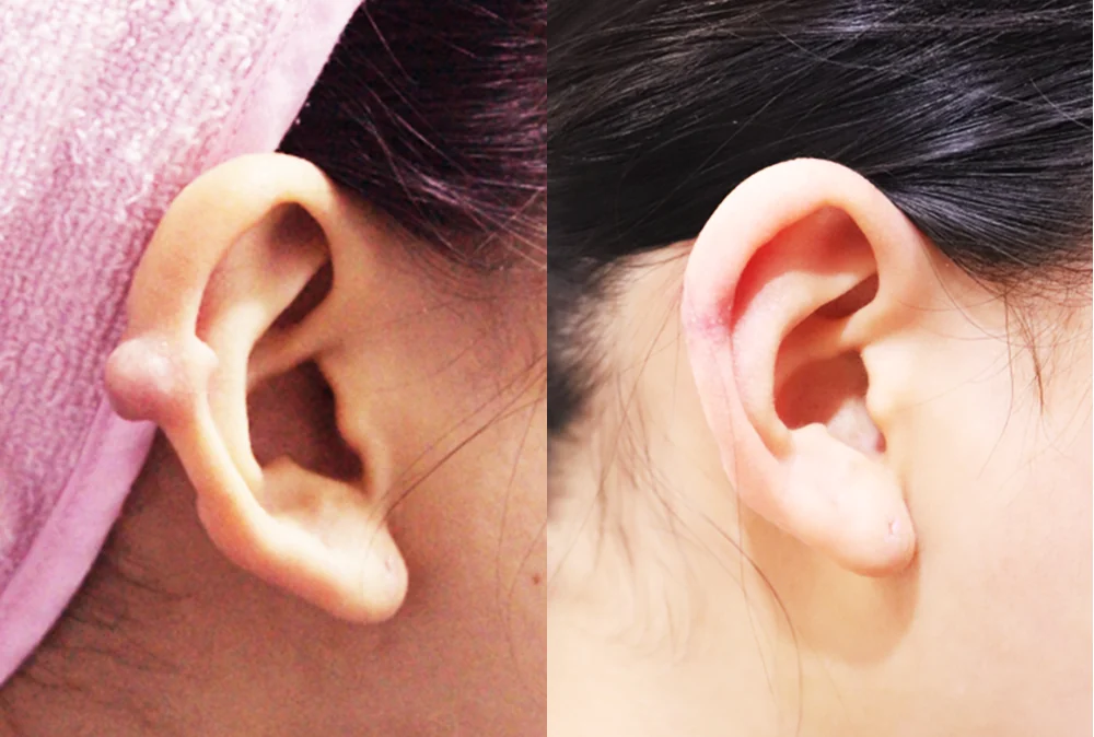
A round lump protruding from the rim of the ear
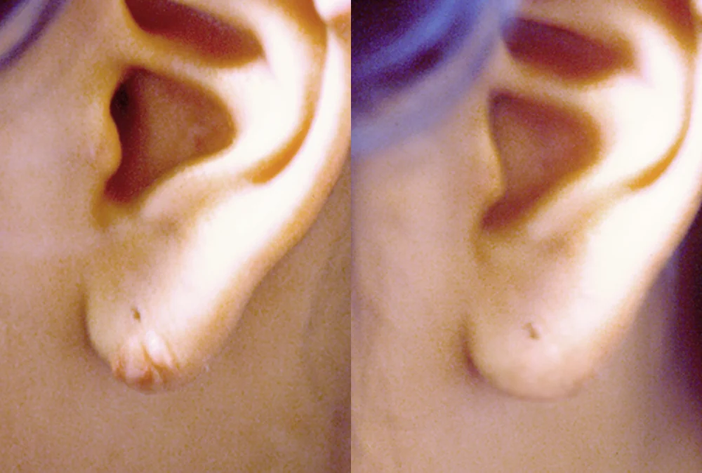
A bulging lump at the tip of the earlobe
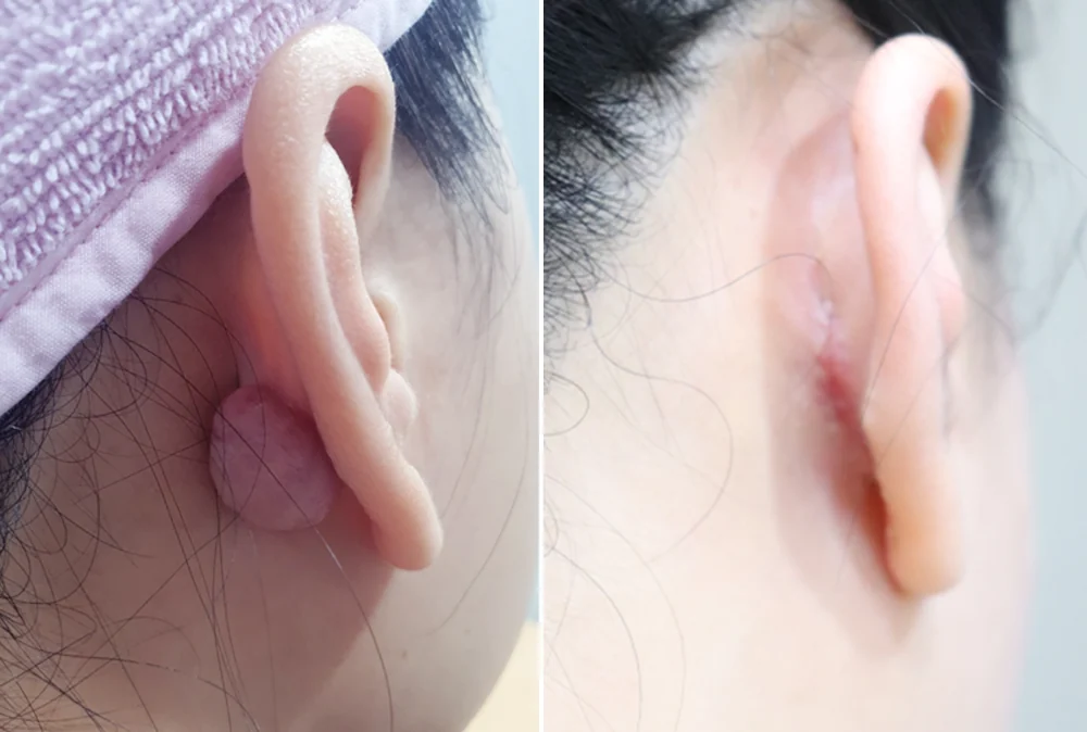
A reddish lump behind the ear
* Results vary from person to person. Side effects are possible, so please decide after a thorough consultation with a doctor.
Frequently asked questions
Can I just squeeze it out at home?
We do not recommend it. Squeezing may empty the contents, but the sac stays and can fill up again, and if the sac ruptures inside the skin the resulting inflammation can leave a larger scar.1 If you have already squeezed or drained it, tell us at consultation so we can check how much of the sac remains.
Will it come back after removal?
If any of the sac remains it can recur, so the sac wall is removed too. Removal does not stop new cysts forming elsewhere, and recurrence at the same site is checked during follow-up.
My cyst is swollen and painful. Can it be removed right away?
When inflammation is active, it is often better to let it settle before removing the cyst.1 If pus has collected and it is very painful, draining it may be needed first. Telling us when the swelling and pain started helps us plan the order.
Will there be a scar?
A short incision line along the skin creases remains. Small lumps without inflammation usually allow a shorter incision, and the remaining line is followed up at our Scar Clinic.
Can a lipoma be removed by liposuction?
Liposuction is used for some lipomas, but compared with removing the lump whole under direct view, it carries a higher risk of leaving fragments that can regrow.4 The choice depends on the size, location and depth of the lipoma.
What to prepare before your consultation
- Where the lump is, when you first noticed it, and whether it has changed in size
- Whether it has ever been swollen, painful, burst, squeezed or drained
- Antibiotics or other treatments you have had for it
- Whether you have ever formed thick, raised scars or keloids
- Medicines you take regularly
Bonita Clinic is on the 3rd floor of 565 Gangnam-daero, Seocho-gu, Seoul, about 100 m on foot from Exit 8 of Nonhyeon Station (Line 7). You can read more on our sebaceous cyst and lipoma removal page. If the incision line left after removal bothers you, see our facial surgical scar page and the guide on when to consider treatment for a surgical scar. For depressed incision scars, see the guide to depressed facial incision scars.
References (6)
Sources that support the explanations in this article. Study findings describe general trends and do not guarantee individual results.
- Zuber TJ. Minimal excision technique for epidermoid (sebaceous) cysts. Am Fam Physician. 2002;65(7):1409-1412.Review: epidermoid cysts are sacs of keratin and lipid (the term 'sebaceous cyst' has fallen out of use), dark keratin plug, causes of the odor, common sites, foreign-body inflammation after rupture, frequent recurrence after drainage alone and prevention by removing the whole sac, waiting about a week for inflammation to settle, and the 2–3 mm minimal excision technique
- Diven DG, Dozier SE, Meyer DJ, Smith EB. Bacteriology of inflamed and uninflamed epidermal inclusion cysts. Arch Dermatol. 1998;134(1):49-51.doi:10.1001/archderm.134.1.49Controlled study (abstract): cultures from 25 inflamed and 25 uninflamed epidermoid cysts showed no significant difference in bacterial number or type, so a red cyst is not necessarily infected
- Lee HE, Yang CH, Chen CH, et al. Comparison of the surgical outcomes of punch incision and elliptical excision in treating epidermal inclusion cysts: a prospective, randomized study. Dermatol Surg. 2006;32(4):520-525.doi:10.1111/j.1524-4725.2006.32105.xRandomized study of 60 patients with non-inflamed cysts (abstract): wound length 0.73 cm with punch incision vs 2.34 cm with elliptical excision, operating time 12.7 vs 21.6 minutes, no difference in recurrence; punch suits 1–2 cm cysts on the face
- Oh SY, Eun S. Minimal One-Quarter Incision and Four-Step (MOQIF) Excision Method for Subcutaneous Lipoma. J Clin Med. 2026;15(6):2448.doi:10.3390/jcm15062448Single-surgeon retrospective series of 82 lipomas: lipoma characteristics, excision as the low-recurrence standard, risk of fragments and recurrence with liposuction or lipolysis, preoperative ultrasound, seroma 11%, hematoma 7.3%, no recurrence at 8.9 months, and the study's limitations
- Johnson CN, Ha AS, Chen E, Davidson D. Lipomatous Soft-tissue Tumors. J Am Acad Orthop Surg. 2018;26(22):779-788.doi:10.5435/JAAOS-D-17-00045Review (abstract): MRI for soft-tissue masses deeper than the fascia or larger than 5 cm in subcutaneous tissue; unplanned sarcoma excisions often follow a presumed diagnosis of lipoma
- Hayes AJ, Nixon IF, Strauss DC, et al. UK guidelines for the management of soft tissue sarcomas. Br J Cancer. 2024;132(1):11-31.doi:10.1038/s41416-024-02674-yUK sarcoma guideline (diagnosis section read): growth, size over 5 cm, deep site and pain were used as referral criteria but did not separate sarcoma well from common lipomas and cysts; most lumps are benign lipomas; ultrasound is a useful first test but operator-dependent, with MRI when uncertain
About this article
This article separates general medical information from Bonita Clinic's own approach. Because the right decision depends on each person's condition, it is not a substitute for a consultation.