KOKorean
 ENEnglish
 JP日本語
JP日本語 CN中文(简体)
 TW中文(繁體)

Keloid or Hypertrophic Scar? How to Tell Them Apart Before Treatment in Seoul
The key difference is the border: a hypertrophic scar stays within the original wound, while a keloid spreads past its edges into the surrounding normal skin. Hypertrophic scars usually rise within weeks, grow for a few months and then settle over years; keloids can appear months or years later, rarely shrink on their own and tend to grow back if simply cut out. Many scars show features of both, so shape, borders, timing, body site and personal risk factors such as family history are weighed together at an examination. The answer decides whether to wait, what to try first, and how much recurrence control to plan around any excision; if you are considering treatment in Seoul, older and current photos of your scar are a good place to start.
Why does it matter whether a raised scar is a keloid or a hypertrophic scar?
When a surgical scar or an old injury heals and then turns red, firm and raised, many people immediately think "keloid". Not every raised scar is a keloid, though. Raised scars fall into two broad groups: hypertrophic scars (scars that have become thick) and keloids.
The two start in the same place. The dermis is the thick layer of skin under the surface, and its deeper part is called the reticular dermis. When a wound reaches that deep layer and inflammation there lasts too long, either kind of scar can form. Shallow wounds that never reach the reticular dermis are described as not causing them.3 The triggers overlap as well: surgery, accidents, burns, piercings, vaccinations, insect bites, acne, folliculitis (inflamed hair follicles), chickenpox and shingles can all start one.3 So "what caused it" is rarely enough to tell them apart.
Why bother telling them apart, then? Because the two scars behave differently over time. A review (a paper that pulls together many studies) points out that misjudging the scar type can lead to the wrong treatment, and even to poor decisions about elective or cosmetic surgery.2 Put simply, the plan depends on whether this is a scar that tends to settle with time, or one that can grow back after it is cut out.
This guide focuses on how the two are told apart (borders, timing, body site), what to do when a scar is unclear, and how the answer changes treatment and recurrence control. The treatments themselves, such as injections, lasers and excision, are covered in our guide to keloid and raised burn scar treatment.
First clue: has the scar spread beyond the original wound?
The single most important difference is the border. A hypertrophic scar thickens only within the area of the original wound. A keloid grows sideways, past the edges of the wound and into the surrounding normal skin.1 A review of studies that compared both scar types side by side reached the same conclusion: sideways growth beyond the wound is the mainstay of a keloid diagnosis.4
To judge this, the scar has to be compared with how long and wide the original wound was. For a surgical scar, the length of the incision and the stitch marks are the reference. For an injury, photos taken right after it happened or when the stitches came out are the reference. If the scar has become clearly wider than the original wound and its ends are pushing out into normal skin, a keloid is the first thing to consider.
The look can differ too. A keloid tends to be a firm, lumpy mass with a shiny surface, sometimes with fine visible blood vessels. Its color ranges from pink to purple, and its edge is sharp but irregular in outline. A hypertrophic scar can look similar, red and raised, but after surgery it usually follows the incision as a line, and after inflammatory spots such as acne it forms small bumps or nodules.2
What you might see in the mirror
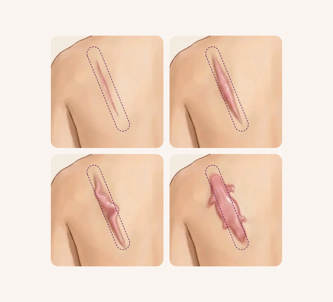
The same spot can look quite different depending on how the scar behaves. Here is what each picture suggests, and what is checked next.
The same scar site, four different pictures
1The line is flat but still red
It may still be maturing, so we watch whether it starts to rise while caring for it
Watch and care
2Raised and thick along the original line
Leans toward a hypertrophic scar; we check whether it stops and settles over months, and how much it pulls
Hypertrophic scar
3Lumpy, and seems to go slightly past the edge
May be an intermediate scar; timing, body site and personal risk factors are weighed together
Intermediate
4Spreading into normal skin like claws
Leans toward a keloid; any excision is planned together with recurrence control
Keloid
A large scar is not automatically a keloid. One report found that a burn that heals in under 10 days carries about a 4% risk of becoming a hypertrophic scar, while a burn that takes 21 days or more to heal carries a risk of 70% or more.3 That is why thick scars after large burns are often hypertrophic scars: wide, but still within the area that was burned. On the other hand, a lump that started from something tiny, such as an acne spot, and has grown well beyond it should raise suspicion of a keloid, whatever its size.
Second clue: when did it appear, and is it still growing?
Time is the next clue. When did the scar start to rise, and is it still getting bigger?
A hypertrophic scar usually starts to rise within 4 to 8 weeks of the injury. It then grows quickly for up to about 6 months and, over a few years, gradually settles and flattens.2 A review of current treatment algorithms describes the same course: it becomes visible a few weeks after the injury, grows for 3 to 6 months, and when risk factors are few, it plateaus and then shrinks on its own.6
A keloid keeps a different timetable. It can appear years after a minor injury, and on the middle of the chest it can even appear without any injury the person remembers. Once it forms, it tends to persist and rarely shrinks on its own.2 The comparative review sums up the difference neatly: hypertrophic scars begin within weeks and grow for months, while keloids can begin months later and have no set end to their growth.4
So a scar that is red and thick two or three months after surgery is not necessarily a keloid. A hypertrophic scar at that stage may simply still be in its growth phase. If, on the other hand, the scar keeps widening after a year, or the itch and pain keep getting worse, keloid features become more likely. Dated photos taken every month or two make these changes much easier to judge, and they are especially useful if you plan to consult a clinic abroad.
Third clue: where on the body is it, and what shape has it taken?
Body site and shape are the third clue. Hypertrophic scars favor places where the skin is stretched a lot, such as the shoulders, neck, the front of the breastbone, knees and ankles. Keloids favor the front of the chest, shoulders, earlobes, upper arms and cheeks, and are uncommon on the eyelids, palms, soles and mucous membranes.2 They also seldom form on the top of the scalp, the front of the lower leg, or the upper eyelid, where the skin is rarely under tension.3
Keloids often take a shape that is typical of their site: a butterfly on the shoulder, a crab's claw on the front of the chest, and a dumbbell on the upper arm. This is explained by the direction in which the skin at each site is pulled. On the front of the chest, for example, the large chest muscle pulls the skin sideways, so keloids there grow horizontally.3 The Japan Scar Workshop consensus (recommendations agreed by experts after reviewing the evidence) likewise notes that both scar types tend to grow in the main direction of skin tension.1
How it started is worth noting too. Keloids can follow obvious wounds such as surgery, but also minor ones such as acne or insect bites.23 A lump that began from such a small trigger and grew large leans strongly toward a keloid.
How do the two scars compare at a glance?
The table below brings the three clues together with symptoms and what tends to happen after treatment. Symptoms differ a little: both scars are commonly itchy, but keloids can also cause significant pain and heightened sensitivity.2 Contracture, where a scar shrinks and stops a joint from straightening fully, is more closely linked to hypertrophic scars.4
Typical hypertrophic scars and keloids compared
| What to look at | Hypertrophic scar | Keloid |
|---|---|---|
| Borders | Stays within the original wound | Spreads into normal skin beyond it |
| Onset | Within weeks of the injury | Can appear months to years later |
| Growth period | Grows for months, then stops | May keep growing |
| Settles on its own | Often, over a few years | Rarely |
| Shape | A line or small nodules | Irregular edges; typical shapes by site |
| Contracture | Can occur near joints | Uncommon |
| After excision alone | A new raised scar is uncommon | Tends to grow back |
One or two rows that do not match do not settle the question on their own. In practice, the whole picture is weighed, along with how the scar changes over time.
What if the scar does not fit neatly into either group?
This is common. Many scars show features of both and sit somewhere in between. The Japan Scar Workshop consensus notes that such intermediate scars often make it hard to say whether a lesion is a keloid or a hypertrophic scar.1 Some experts see the two as opposite ends of the same disorder, with a range of intermediate scars between them; which end a scar lands on depends on how strong and how long-lasting the inflammation in the reticular dermis is.36 Others argue that sideways growth is a real, defining difference, and that the two should still be treated as separate conditions for now.4
A scoring scale
The Japan Scar Workshop uses a scoring scale called JSS 2015. It scores how raised the scar is, whether it has spread sideways, its shape and the redness around it, and adds risk factors such as an early age of onset. A total of 5 or less suggests a mature (settled) scar, 6 to 15 a scar with strong hypertrophic features, and 16 or more a scar with strong keloid features.1 According to the consensus, scars scoring 6 to 15 tend to respond well to treatment, while those scoring 16 or more tend to resist treatment or come back, and are better managed where several treatments can be combined.1 In other words, the score is less about attaching a label and more about estimating how stubborn a scar is likely to be.
Can a biopsy or ultrasound settle it?
Not always. Under the microscope, keloids contain thick, glassy, evenly stained fibers called keloidal collagen, while hypertrophic scars contain nodules of tightly packed fiber bundles. Keloids can contain those nodules too, however, and there is no agreed cut-off that separates the two. As a result, the diagnosis made by eye and the diagnosis made under the microscope do not always agree.1
The numbers make the point. In a study comparing tissue from 40 keloids and 10 hypertrophic scars, keloidal collagen was found in only 55% of the keloid samples. A marker often linked to hypertrophic scars (alpha-smooth muscle actin) was found in 70% of hypertrophic scars and 45% of keloids, so it could not separate them either.5 The same study did confirm that keloidal collagen is useful for diagnosis. It also reported that even when no keloidal collagen is found, a few features, such as a tongue-like edge pushing in under normal-looking skin, point toward a keloid.5 No blood or tissue marker that reliably tells the two apart has been found yet.1
Ultrasound, including elastography (ultrasound that shows how stiff tissue is), can measure a scar's thickness and firmness and track changes during treatment. Imaging alone, however, cannot reliably tell a keloid from a hypertrophic scar.1 In the end, the examination is central: the scar's relation to the original wound, when it appeared and how fast it grew, its site, and the person's own risk factors, all considered together.
What does the edge of the scar look like?
- Stays inside the original lineLeans hypertrophicWatch whether it stops and settles over months
- Spreads into normal skinLeans keloidEven more so if it began from a tiny wound
- Seems slightly past itIntermediateTiming, site and personal risk weighed together
- Grows fast or looks unusualPossibly another growthConsider a biopsy first
The name you were given may depend on where you were seen
Keloids are common in people of African descent (5 to 10%), less common in Asians (0.1 to 1%), and rare in people of European or North American descent (under 0.1%).6 One review author, describing an impression formed at international scar conferences, suggests this shapes how doctors label scars: where hypertrophic scars dominate, scars with even slight spreading may be called keloids, while where keloids are common, scars may be called hypertrophic unless they grow quickly.6 If you were told one name at home and another elsewhere, the most useful step is to look again at the scar's borders and its course over time.
Could a "keloid" actually be a different growth?
Rarely, yes. In one analysis of 378 scars thought to be hypertrophic scars or keloids, 1.06% turned out to be other conditions,6 roughly 1 in 100. The Japan Scar Workshop consensus lists skin growths that can look like keloids or hypertrophic scars. Benign (non-spreading) ones include pseudolymphoma, mixed tumor of the skin, xanthogranuloma, leiomyoma and dermatofibroma. If one of these is suspected, a biopsy (removing a small piece of tissue to examine under a microscope) should be considered before treatment.1
Some malignant growths can also resemble keloids or hypertrophic scars: dermatofibrosarcoma protuberans, cutaneous squamous cell carcinoma and amelanotic (non-pigmented) melanoma. If a malignant growth is suspected, for example because a lump is growing rapidly, a biopsy is essential. Ultrasound, CT or MRI beforehand helps map the lesion for that biopsy.1
So in these situations, keloid treatment is not started straight away; the diagnosis is confirmed first:
- A lump that appeared without a clear wound and is growing quickly over a short time
- Features on examination that differ from a typical keloid or hypertrophic scar, raising suspicion of another growth
How does the diagnosis change the treatment plan?
The answer changes the plan because the two scars are heading in different directions.
Hypertrophic scars tend to settle with time, so surgery is seldom needed unless the scar contracts and restricts a joint. Conservative care (care without surgery), such as pressure, silicone sheets, tape or injections, can speed up settling and ease itch and pain.6 When contracture is severe, surgery to release it comes first. When it is mild, the options are to remove the scar completely or to combine several non-surgical treatments.6 After a hypertrophic scar is excised, a new hypertrophic scar in the same place is uncommon.2
Keloids rarely shrink on their own and tend to grow back if they are simply cut out.2 The algorithm review reports recurrence of 45% or more after excision alone, and advises that complete removal of a keloid should always be combined with an added (adjuvant) treatment such as radiotherapy.6 Small, single keloids can be treated by excision with an added treatment, or by several non-surgical treatments combined. For large or multiple keloids, surgery that reduces their bulk or number is one option.6
Every treatment also has side effects and limits. Steroid injections into the scar (an anti-inflammatory medicine) can be painful and can cause body-wide side effects such as irregular periods. The skin at the injection site can also become thinner, show small widened blood vessels, or turn lighter in color.6 Surgery such as excision can be followed by bleeding, infection or inflammation. Added treatment does not remove the risk of recurrence either: in a meta-analysis cited by the same review, which pooled 72 studies, keloids recurred in 22% after surgery combined with radiotherapy.6 That is roughly one in five.
In short, treatment of a hypertrophic scar works with time and helps it settle, while treatment of a keloid holds back its growth and manages recurrence over the long run. Either way, long-term follow-up after treatment is recommended.6 The evidence and limits of each method, from injections and tape to lasers and excision, are covered in our guide to keloid and raised burn scar treatment, and the timing of surgical scar treatment in our guide to when to treat a surgical scar.
What makes these scars more likely to form, worsen or come back?
Whether a keloid or hypertrophic scar grows back or worsens depends on local conditions at the scar, the state of the body as a whole, inherited factors and lifestyle, all acting together.6
- Tension: the more often the skin is stretched, the more both scar types tend to worsen. Scars in manual workers and athletes who repeat the same movements tend to be stubborn and need a longer treatment period.1
- Repeated injury and inflammation: putting earrings in and taking them out again re-injures a piercing hole over a long period, which greatly raises the risk of earlobe keloids. Repeated infection of piercing holes, acne and folliculitis also raise the risk.3
- The whole body: adolescence and pregnancy appear to be linked to higher risk.3 In a study of 304 people with keloids, higher blood pressure was linked to larger and more numerous keloids.3 Conditions that raise inflammatory substances in the blood can also make these scars worse.1
- Inheritance: some people have a family history of keloids, and people with darker skin are reported to be about 15 times more likely to develop these scars. A genome study in Japanese people identified genetic variant sites associated with keloids.3
The algorithm review advises that people with these risk factors be managed with particular care during and after any surgery.6 And if you have had a keloid and are planning another operation, a cosmetic procedure or a piercing, tell the doctor first. Misjudging the scar type can lead to poor decisions about cosmetic surgery.2
How we approach it at Bonita Clinic
At Bonita Clinic, we treat keloids and hypertrophic scars that formed after accidents or procedural complications, surgery, caesarean sections, and implant or filler removal. Treatment is an in-clinic procedure, with excision when needed, under local anesthesia, and you go home the same day. Stitches are used only if excision is done. Recovery time and the number of visits depend on the extent of treatment and your condition. You can consult even before a wound has fully healed.
Treatment options you can discuss in Seoul
Bonita Clinic opened in 1995 and has stayed at the same location since, where Dr. Kwang Sub Jung, a plastic surgeon board-certified in Brazil, sees patients. For raised scars, the options discussed in consultation are the ones on our treatment page:
- Injection treatment for red, firm, raised keloids, repeated at intervals
- Excision with low-tension suturing, followed by recurrence management
- Combined lasers for remaining redness, pigmentation and texture
- For tight, hardened burn scars, release of the tight tissue and fat stem cell treatment
Which of these fits, and in what order, depends first on whether your scar behaves like a keloid or a hypertrophic scar, and that is confirmed by examination.
Why the plan and cost differ from scar to scar
A flat red line, a thick hypertrophic scar along an incision and a spreading keloid on the shoulder are very different projects. It is the scar's behavior and the extent of the work, rather than its name alone, that shape the plan and the cost.
- Keloid, hypertrophic or in between
- Size, number and body site
- Injections, excision or both
- Laser for remaining redness or texture
- Number of sessions and spacing
- Earlier treatment and recurrences
Final plan and quote after an in-person exam
A final estimate is given only after an in-person examination, once the scar type, the methods and the likely number of sessions are clear. Photos help narrow down the options, but the scope is set in person.
What photos can show, and what needs an in-person exam
Photos are a genuinely useful first step for this question, because the key clue, whether a scar has spread beyond the original wound, can often be seen by comparing old and new pictures. Firmness, tightness and symptoms, however, cannot be judged from a screen.
Photos can show
- Location, size and color
- Spread past the original wound (with older photos)
- Shape and number of scars
- Change over time (dated photos)
Needs an in-person exam
- Firmness and thickness by touch
- Tightness and joint movement
- Itch, pain and tenderness
- Signs that call for a biopsy
- Surrounding skin and other scars
Helpful photos: the whole scar and a close-up in daylight without filters, with a ruler or coin beside it for scale if you can, plus any older photos showing the original wound or stitches. Add dated photos if the scar has changed over the past months. You can send us a message with your photos, the cause and date of the original wound, and the treatments you have had so far.
Planning your visit and follow-up after you return home
What is confirmed in advance is limited to the treatment information above: a procedure with excision if needed, local anesthesia, same-day discharge, and stitches only if excised. Injections are repeated at intervals, and any excision is planned separately, so how many times you come to Seoul and how long you stay are decided with you at consultation according to the scar type, size and site.
Both scar types call for long follow-up after treatment, often well beyond the treatment visits themselves.6 Before you leave, ask which signs to watch for, such as new itch, redness at the edge, or regrowth past the line. Keep taking dated photos from the same angle so that changes can be compared with your consultation photos. If a treated area becomes painful, red and hot, or starts to discharge after you return home, see a local doctor promptly.
Real treatment cases
These cases appear on Bonita Clinic's burn and keloid treatment page. Photos alone cannot show whether each scar was a keloid or a hypertrophic scar, so only what is visible is described. Results vary from person to person.
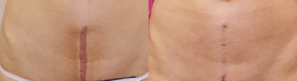
Vertical surgical scar on the lower abdomen, red and raised along the line
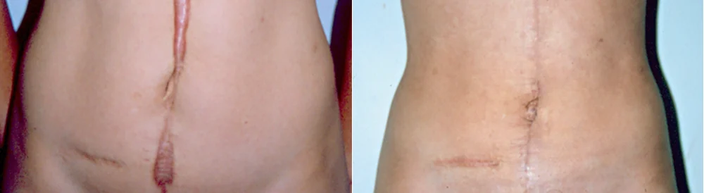
Vertical midline abdominal scar, thick and lumpy, with a horizontal scar below
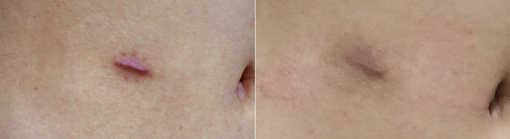
Short scar beside the navel, red and raised
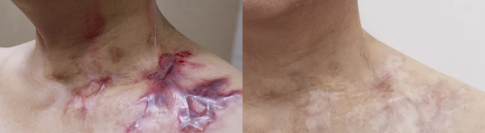
Burn scar below the neck and over the collarbone, with raised red bands branching out
* Results vary from person to person. Side effects are possible, so please decide after a thorough consultation with a doctor.
Frequently asked questions
Can a photo show whether my scar is a keloid?
A photo shows the location, size and color, and with older photos it can suggest whether the scar has become wider than the original wound. Firmness, tightness, itch and pain, and the possibility of a different growth, can only be checked in person. Because many scars sit between the two types,1 photos are used as a starting point and the diagnosis is made by examination.
Can a hypertrophic scar turn into a keloid later?
Some experts see the two as opposite ends of one disorder, with intermediate scars in between.6 So even a scar that first stayed inside the wound is reassessed as having keloid features if it later starts to spread past the edge, and the plan is changed accordingly. While a scar is still growing, dated photos make that judgement much easier.
I have had a keloid. Can I still have other surgery or a piercing?
Having had a keloid does not mean every operation must be avoided. Misjudging the scar type can lead to poor decisions about cosmetic surgery, though,2 so tell your doctor where and when the keloid formed and how it was treated. With piercings, the repeated irritation of putting earrings in and out, and infection, are recognized factors that raise the risk of earlobe keloids.3 For surgery at keloid-prone sites such as the chest, shoulders or upper arms, the incision site and aftercare are worth discussing in advance.
My scar itches and hurts. Does that mean it is a keloid?
Itch is common with both scar types. Keloids can also cause significant pain and heightened sensitivity.2 A growing keloid shows strong inflammation at the edge where it meets normal skin.1 If the itch and pain are getting worse while the edge turns redder and wider, the scar may be growing, and it is worth having it examined.
Are keloids inherited?
Some people with keloids have relatives with keloids too, so inherited factors are thought to play a part. A genome study in Japanese people found genetic variant sites associated with keloids, but much about the responsible genes is still unknown.3 If keloids run in your family, mention it before an injury is treated or an operation is planned, so that your tendency to scar can be factored into the aftercare plan.
Before your consultation
- The cause of the scar (surgery, injury, burn, acne, piercing) and the date
- Photos that show the size of the original wound (right after the injury or when stitches came out) and current photos
- When the scar started to rise, and whether it is still growing
- Itch or pain, and when it gets worse
- Treatments so far: number and date of the last injections, tape or silicone, laser, excision, radiotherapy
- Family history of keloids, high blood pressure, pregnancy plans
Bonita Clinic is on the 3rd floor of 565 Gangnam-daero, Seocho-gu, Seoul, about 100 m on foot from Exit 8 of Nonhyeon Station (Line 7). You can read more on our Burn & Keloid Treatment page. For settled burn scars, see Burn Scars and our guide to settled burn scars; for scars left after facial cosmetic surgery, see Face Surgery Scars.
References (6)
Sources that support the explanations in this article. Study findings describe general trends and do not guarantee individual results.
- Ogawa R, Akita S, Akaishi S, et al. Diagnosis and Treatment of Keloids and Hypertrophic Scars-Japan Scar Workshop Consensus Document 2018. Burns Trauma. 2019;7:39.doi:10.1186/s41038-019-0175-yJapan Scar Workshop consensus: hypertrophic scars stay within the wound while keloids grow sideways beyond it; intermediate lesions are common; JSS 2015 scores (5 or less mature, 6–15 hypertrophic features, 16 or more keloid features); no distinguishing biomarker; keloidal collagen versus nodules without a clear cut-off; inflammation at the growing edge; limits of ultrasound and elastography; benign and malignant look-alikes and biopsy; tension, pregnancy, hypertension, manual workers and athletes
- Gauglitz GG, Korting HC, Pavicic T, et al. Hypertrophic Scarring and Keloids: Pathomechanisms and Current and Emerging Treatment Strategies. Mol Med. 2011;17(1-2):113-125.doi:10.2119/molmed.2009.00153Review: hypertrophic scars appear within 4–8 weeks, grow for up to 6 months and settle over years, while keloids can appear years after minor injury or spontaneously on the chest and do not regress; appearance and pain; typical sites; keloids recur after excision whereas new hypertrophic scars are rare; misidentification leads to inappropriate treatment and cosmetic-surgery decisions
- Ogawa R. Keloid and Hypertrophic Scars Are the Result of Chronic Inflammation in the Reticular Dermis. Int J Mol Sci. 2017;18(3):606.doi:10.3390/ijms18030606Review: these scars follow injury reaching the reticular dermis (surgery, piercing, acne and more); burn healing time and hypertrophic scar risk (4% under 10 days, 70% or more at 21 days or longer); site-specific keloid shapes and direction of tension; rare sites; earrings and infection as local factors; adolescence, pregnancy and hypertension (304 patients); family history, skin color and genetic variants; the view that both lie on one spectrum
- Limandjaja GC, Niessen FB, Scheper RJ, Gibbs S. Hypertrophic scars and keloids: Overview of the evidence and practical guide for differentiating between these abnormal scars. Exp Dermatol. 2021;30(1):146-161.doi:10.1111/exd.14121Review of studies comparing both scar types within the same study: sideways growth is the mainstay of keloid diagnosis; differences in onset (weeks versus months), growth period, spontaneous regression and contracture; tissue differences are mostly a matter of degree, yet treating them as separate entities remains appropriate
- Lee JY, Yang CC, Chao SC, Wong TW. Histopathological differential diagnosis of keloid and hypertrophic scar. Am J Dermatopathol. 2004;26(5):379-384.doi:10.1097/00000372-200410000-00006Tissue comparison of 40 keloids and 10 hypertrophic scars (abstract): keloidal collagen was present in only 55% of keloids, and alpha-smooth muscle actin was seen in 70% of hypertrophic scars and 45% of keloids, so it did not separate them; the study still confirmed the diagnostic value of keloidal collagen, and found that even without it, features such as a tongue-like advancing edge favor a keloid
- Ogawa R. The Most Current Algorithms for the Treatment and Prevention of Hypertrophic Scars and Keloids: A 2020 Update of the Algorithms Published 10 Years Ago. Plast Reconstr Surg. 2022;149(1):79e-94e.doi:10.1097/PRS.0000000000008667Algorithm review: hypertrophic scars appear after weeks, grow 3–6 months and regress; surgery is rarely needed without contracture; keloids recur in 45% or more after excision alone, so adjuvant therapy is advised (22% recurrence with surgery plus radiotherapy in a 72-study meta-analysis); choices by keloid size and number; drawbacks of steroid injection (pain, menstrual irregularity, thin skin, widened capillaries, lighter skin); 1.06% of 378 presumed scars were other diseases; extra care around surgery for at-risk people; frequency by ancestry and regional labelling; long-term follow-up
About this article
This article separates general medical information from Bonita Clinic's own approach. Because the right decision depends on each person's condition, it is not a substitute for a consultation.